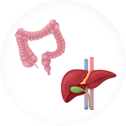


Obesogenic NASH models featuring all the major indicators of NAFLD to moderate NASH, including dyslipidemia, inflammation, ballooning and robust fibrosis
Pharmacologic responses
Reference compounds (OCA, Elafibrinor, Semaglutide) in our high fat diet + modified high fat diet (HFD+mHFD) NASH mouse model match the responses of these experimental drugs in clinical trials
Histology and biomarkers
Demonstrate clinically relevant levels of disease phenotype & scores
Includes steatosis, ballooning, inflammation, mallory body and fibrosis
Metabolic profiles
Similarly altered obesity markers as does human NASH, including insulin, glucose, cholesterol, triglycerides and free fatty acid
Mechanisms of action
Drugs targeting diverse biological pathways and processes
Speed
High fat diet + modified high fat diet (HFD + mHFD) mouse model with shorter induction period between 12 – 16 weeks
High fat, high cholesterol + Fructose (HFC+F) hamster models with induction period between 8 – 12 weeks
Comprehensive
5 different NASH disease models
Multiple induction methods
Obesogenic (HFD, mHFD, AMLN)
Chemical induction (CCl4, AMLN+CCl4)
Nutrient deficient (0.1MCD)
Collaborative & Responsive
Group leaders & study directors provide rapid feedbacks and consultations to your preclinical model studies, including model selection, study design and data interpretation
NAFLD/NASH Models & in Vitro Assays
Nutrient-deficient NASH Models
SD rat or C57bl mice fed on MCD diet for 2-3 weeks
SD rat or C57bl mice fed on 0.1MCD diet for 2-6 weeks
Chemically induced NASH Models
AMLN/HFD + CCl4 in C57bl mice, 12+4 weeks
STZ induced STAM NASH mode, 6+8 weeks
Obesogenic dietary NASH Models
HFD + modified HFD/WD in C57bl mice, 12-16 weeks
Genetic NASH Models
ob/ob mice with AMLN diet, 8-12 weeks
Hamster NASH Model
Golden Syrian hamsters
High fat & high cholesterol (HFC) plus fructose, 8-12 weeks
Fatty Liver
Liver weight
Liver Function: ALT, AST
Lipid panel (liver & serum): TC, TG, FFA
Insulin resistance: Insulin, BG
Histology: HE
Hepatic Fibrosis markers
Collagen type 1a, 3a, α-1 Procollage
SMA-a, TGF-b
Hydroxyproline
Histology Sirius Red staining
Hepatic Inflammation markers
TNF-a levels
Mononuclear cell infiltration
Oxidative Stress
Reactive oxygen species
Hydroxynonenal (HNE) level
Preclinical Models for MASH Disease
MASH Mouse and Animal Models Development and Testing